Procedures
Fillings
Small to medium-sized cavities are repaired with fillings. Typically, this size cavity lesion is diagnosed on a dental x-ray and/or our experienced dentists during our exam. By the time we can see a large cavity or hole in a baby tooth especially, often this size of a cavity may need more extensive treatment such as a crown. In our office, we used tooth-colored fillings. The procedure involves freezing or numbing the affected tooth, removal of the decayed/cavitated, or unhealthy tooth structure, and filling the hole with the tooth-colored filling material. A filling is a conservative way to treat a cavity. Leaving decay unchecked can result in the cavity progressing and needing more invasive, expensive treatment. Baby tooth cavities can progress very quickly due to the structural nature of these small teeth. We encourage you to keep up with your child’s preventative dental checkups, where we can detect cavities at their earliest stages!
Sealants
A sealant is a protective coating that is applied to the chewing surfaces (grooves) and deep anatomy (pits and fissures), typically on the permanent molar teeth. This sealant acts as a barrier to food, plaque, and acid, thus protecting these more vulnerable areas of the teeth. Our doctors will evaluate your child for their particular risk for cavities on these areas, and whether sealants would be indicated.
Nerve Treatments
When a cavity gets moderate to deep in size, it can affect and communicate with the pulp tissue of the tooth-which is what we call the very center of the tooth where the nerves, tissue, and blood vessels are found in both primary and permanent teeth. This requires us to remove the affected pulp tissue in order to prevent infection and further deterioration/disease of the tooth and its supporting structures.
There are 2 types of primary tooth nerve treatments:
- Pulpotomy: If the disease has not advanced into the root of the tooth, the pulp within the crown of the tooth is removed, and a medicament is placed over the remaining healthy tissue in the roots of the tooth. This dressing will help to keep the remaining pulp tissue and the tooth healthy.
- Pulpectomy: If the disease has advanced into the root of the tooth, the entire pulp or nerve tissue needs to be removed and the canals of the tooth cleaned. A resorbable medicated dressing is placed that will dissolve with the root of the tooth when the permanent successor tooth is advancing and growing into the mouth.
Once a baby tooth needs either a pulpotomy, or a pulpectomy, it is necessary for that tooth to have a crown restoration placed, in order to provide a good seal, structural support, and thus ensure the highest chance for a successful treatment outcome.
Permanent Teeth have several types of nerve treatments as well:
- Indirect Pulp Cap: When removal of a cavity is very close to the nerve of the tooth, but no exposure has occurred, placing a medicated layer close to the nerve can help the tooth to heal and form a reparative layer to further insulate the nerve and prevent inflammation and the possible deterioration of the nerve. If a tooth is chipped and the same scenario occurs due to dental trauma, in that the nerve is very near exposure, placing this type of indirect pulp cap can also help heal the nerve in the same manner.
- Direct Pulp Cap: When removal of a cavity results in a very small nerve exposure on an otherwise asymptomatic tooth, or on an immature permanent tooth, placing a medicated layer directly onto the nerve of the tooth can help the tooth to heal and form a reparative layer to insulate the nerve, and prevent inflammation and deterioration of the nerve. The same scenario can occur if a tooth is chipped due to dental trauma, and the nerve is exposed.
- Root canal: When removal of a cavity results in a nerve exposure on a mature permanent tooth, complete removal of the nerve is necessary, with thorough cleansing and shaping of the canals, and placing of a permanent filling material within the canals. If this type of procedure is required, our doctors and team will refer you to one of our local endodontists, who specialize in this area of dentistry. Permanent teeth that require a root canal often will require a crown, especially if the nerve treatment was due to a large cavity or fracture.
Crown:
A dental crown (sometimes called a cap) is a tooth-shaped restoration that completely covers or surrounds the “crown” or the portion of the tooth visible in the mouth. It protects and supports the tooth, preventing further cavities and breakdowns from occurring.
In primary or baby teeth with moderate to deep/large cavities, or significant wear, a filling is no longer sufficient to restore the tooth. If your child has a very high cavity rate, a crown might also be chosen to prevent an additional breakdown on intact surfaces that are compromised or anticipated to need treatment. For children, crowns come in pre-fabricated sizes, so that the crown procedure can be completed in one visit.
Baby tooth crowns come in two varieties:
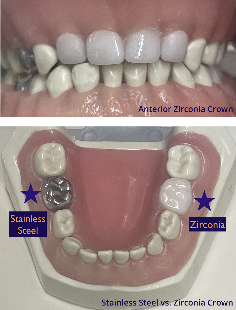
- Stainless Steel crowns: made of a safe, stainless steel alloy, which makes this type of crown somewhat adjustable due to its ability to be bent and trimmed. These are very strong restorations that are efficient to place during a procedure. Our patients refer to them as “superhero” crowns! Permanent teeth that need a temporary crown until your child is fully grown and ready for an adult crown, are made from stainless steel.
- Zirconia (tooth-colored) porcelain crowns: made of a rigid, tooth-colored zirconium oxide material that is 100% metal free. These crowns are very esthetic, providing a natural looking option to cap or crown a baby tooth. Due to the rigid, non-flexing properties of the zirconia material, they do require a very technique sensitive preparation, and exact fit. Our doctors are Sprig certified providers of these crowns for anterior and posterior baby teeth.
Our doctors can help you to choose which crown is appropriate for your child’s teeth.
Extractions:
Teeth sometimes need to be removed due to severe decay, trauma, malposition, or interference of a baby tooth with the eruption of a permanent tooth, failure of a baby tooth to exfoliate/fall out completely. If extraction is indicated, our team will discuss whether a space maintainer that helps to keep the adjacent or developing teeth from shifting and creating positional issues later, is recommended. Our doctors will make sure to freeze/numb the area to ensure your child is comfortable.
In some cases where extractions may require surgery of the gums, or a permanent tooth needs to be removed, our team will help to refer you to a local oral surgeon that specializes in this area.
Space Maintainers:
Baby teeth are extremely important for many reasons. They not only help with essential functions including chewing and speech, but also play a role in growth and development of the jawbones and muscles and hold space and guide the developing adult teeth into position.
When baby teeth are lost prematurely, before the permanent successor is/are ready to erupt, in order to prevent shifting and loss of space, it is important to place a space maintainer. Space maintainers are custom-fit metal devices that are anchored on a neighboring tooth and “hold” the space of the lost baby tooth to prevent shifting and loss of space from occurring, which might later result in an impacted adult tooth and/or the need for orthodontic correction.
Children do well with adapting to the presence of the space maintainer in their mouths. These are generally cemented into place, and remain until a dentist removes them with a special instrument at the appropriate stage of development. Our team will coach you on proper care and maintenance of the appliance. Avoiding very sticky foods such as taffy or caramels that might dislodge the spacer is important. If it does become loose or fall out, please contact our office. It is important to maintain your child’s regular twice-yearly checkups so we can adjust and or monitor the space maintainer.
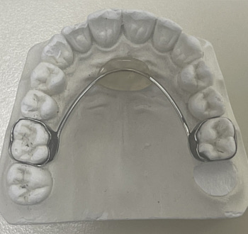 Nance/bilateral space maintainer
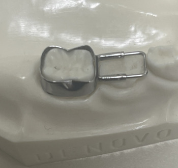 Band and loop/unilateral space maintainer
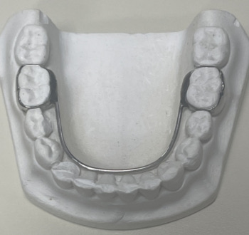 Lower lingual holding arch/bilateral space maintainer
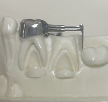 Distal shoe/unilateral space maintainer
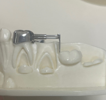 Distal shoe space maintainer
Silver Diamine Fluoride:
Silver diamine fluoride, or SDF, is an antimicrobial treatment that can help to arrest/stop the progression of tooth decay and reduce dental sensitivity associated with cavities and enamel defects. It has been used extensively in other countries for decades with many studies proving it’s efficacy and safety, and it is FDA approved. The free silver ion from SDF is bactericidal. When applied to a tooth with a cavity, the active lesion becomes inactive, and the risk of future decay is also reduced. SDF turns the cavity black/very dark in color, forming a scar. The scarred area is remineralized and hardened from the fluoride in SDF, and more acid/abrasion resistant. The scar is also reduced in sensitivity to temperatures and sweets.
When do we use SDF?
- Halting cavities from progressing if definitively restoring/fixing teeth is not possible or recommended currently due to one of several possible factors such as:
- Patient age, maturity, or special health needs do not allow for a definitive treatment currently
- The tooth is not fully erupted or accessible in the current position to provide final treatment, and will require staged treatment over time
- Reducing sensitivity in permanent molars with hypoplastic enamel so future restorative treatment is comfortable/tolerable.
Icon Resin Infiltration (ICON):
What is ICON? Resin infiltration is a minimally invasive restorative treatment for white-spot lesions (WSLs) and certain congenital hypocalcified enamel lesions. White spot lesions are enamel porosities caused by demineralization of the tooth enamel due to poor oral hygiene/plaque accumulation, and acids from the bacteria and/or in the diet breaking down the surface of the enamel. This results in the white/chalky appearance of the demineralized enamel. Hypocalcified lesions are congenital enamel defects often of unknown origin, but in some cases caused by prior trauma to the baby tooth in the area, which has affected the enamel formation of the adult tooth. In the ICON procedure, the tooth enamel is etched to open the tubules/porosities within the tooth and allow access to the internal structure where the enamel has been disrupted. The unfilled resin is then applied to the area so that it infiltrates the structure and mimics the optic properties of the natural tooth structure, thereby camouflaging/fading the previous noticeable white spot. There is no removal of structure, and no need for numbing/anesthetic. The results have been shown to last for 2+ years. We have had several patients with stable results for many years beyond this! We cannot guarantee the spot will disappear completely, but in most cases, it provides a significant esthetic improvement in one visit, without any invasive or irreversible procedure. Our doctors would be happy to advise if ICON might be helpful in the case of an esthetic white spot lesion on your child’s tooth.
![]()
